Search Close Search
Search Close Search
Page Menu

Awardees

  • Parikh.jpg

    Modulating the immunosuppressive pancreas tumor microenvironment through intratumoral delivery of cytokine-encoding mRNAs

    Pancreatic ductal adenocarcinoma (PDAC) is an extremely aggressive cancer, with a mere 13% average 5-year survival rate. Conventional chemotherapy and immunotherapy regimens have limited effectiveness, and surgical resection of the tumor is not viable for the majority of PDAC patients diagnosed with advanced metastatic disease. Our previous research has shown therapeutic induction of cellular senescence with RAS pathway targeting therapies can induce cytokine and chemokine production through the senescence-associated secretory phenotype (SASP) that can effectively activate anti-tumor T cell immunity and responses to anti-PD-1 immune checkpoint blockade. To build on these findings and overcome limitations associated with senescence-inducing therapy toxicity, here we leveraged mRNA technology to deliver specific SASP cytokines and chemokines we have found to stimulate immune responses into the suppressive PDAC TME as an immune oncology platform. We created an in vitro transcription pipeline for mRNA production and have successfully demonstrated delivery of multiple mRNAs simultaneously into the TME of transplanted and autochthonous PDAC mouse models, stimulating the local production of cytokines normally absent in PDAC. We further characterized a novel combination of five cytokines and chemokines that can effectively recruit and activate both innate and adaptive immune responses against PDAC. Considering the recent success of off-the-shelf neoantigen mRNA vaccines in early human PDAC patient trials, we have now developed mRNAs that encode PDAC-specific antigens. By integrating our cytokine mRNA platform with antigen mRNAs, we observe a significant increase in intratumoral antigen-presenting dendritic cell populations, leading to substantially enhanced T-cell-mediated anti-tumor immune response. Long-term studies in autochthonous PDAC mouse models demonstrate that combining cytokine and antigen mRNAs can completely eradicate tumors in select cases, resulting in significantly extended survival. Overall, this study not only unveils pivotal insights into the cytokines absent in PDAC that are crucial for promoting anti-tumor immunity but also pioneers an innovative immunotherapy approach.

    Read more
  • Zvornicanin.jpg

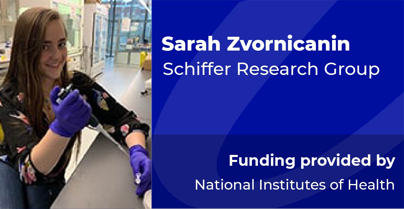
    Structure-based Antiviral Design against HTLV-1 Protease

    Human T-cell leukemia virus type-1 (HTLV-1) is an oncogenic human retrovirus affecting over 20 million people worldwide. HTLV-1 infection can cause adult T-cell lymphoma (ATL) and other serious inflammatory diseases. Estimates report that 5-10% of HTLV-1 infected patients will develop a serious condition such as ATL, which has poor 4-year survival and high relapse rates. HTLV-1 has persistent infection rates across the globe and reaches up to 45% prevalence in certain communities. Despite this impact on human health, there are no direct-acting antivirals (DAAs) or vaccines against HTLV-1. HIV-1 and HTLV-1 are from the same viral family and encode for a homodimeric aspartyl protease crucial for cleavage of functional proteins from viral polyproteins. The activity of HIV-1 and HTLV-1 protease is essential to their viral life cycles. The Schiffer laboratory has extensive experience with viral protease crystallography and inhibition, especially with viral proteases for HIV-1, HCV NS3/4A, and SARS-CoV-2 main protease. This expertise uniquely positions me to design, synthesize, and characterize potent, resistance-thwarting protease inhibitors against HTLV-1 protease. Resistance-preventing DAA design is essential because of the selective pressure applied during DAA treatment. An ideal and proven strategy for developing a highly potent and resistance-preventing viral protease inhibitor is to target the active site through rational design using the substrate envelope. The substrate envelope for HTLV-1 protease has not been characterized and we lack a detailed understanding of the protease substrate specificity. I hypothesize that by translating strategies from our design of HIV-1 protease inhibitors, namely characterizing HTLV-1 protease’s substrate specificity, I can design potent and resistance-preventing DAAs for HTLV-1 protease. Aim 1: Characterize the structural basis for HTLV-1 protease substrate specificity. HTLV-1 protease cleaves six substrates by recognizing cleavage sites between individual proteins of the viral polyprotein. I will investigate the molecular basis of this recognition underlying protease specificity by determining cocrystal structures of the protease with bound substrates. The conserved volume inhabited by the substrates will define the substrate envelope and inform inhibitor design for HTLV-1 protease. Aim 2: Rationally design, synthesize, and characterize inhibitors of HTLV-1 protease to optimize potency. HIV-1 and HTLV-1 proteases share an active site amino acid sequence identity of 45% and high structural similarity. Therefore, I will begin inhibitor design by testing a selection of our in-house HIV-1 protease inhibitors, which have already shown low (1 µM) to moderate (30 nM) potency against HTLV-1 protease. I will combine experimental inhibition assays with cocrystal structure analysis to identify lead compounds for inhibitor design. I will leverage substrate specificity of the protease by moving inhibitor design towards compounds that mimic the shape of substrates, leveraging the substrate envelope (Aim 1), and the interactions between protease and substrate. I aim to produce novel, highly potent (sub-nM) inhibitors that will be promising DAAs for further investigation against HTLV-1.

    Read more
  • Allen.jpg

    Examining the Role of a Pathogenic HTT Isoform, HTT1a, in Somatic Expansion and RNA Aggregation in Huntington's Disease

    Huntington’s Disease – a devastating neurodegenerative condition – is caused by a defect in the Huntingtin gene, resulting in the production of alternative forms of Huntingtin mRNA and protein. This proposal will use small RNA drugs to reduce alternative forms of Huntingtin in mouse models of Huntington’s disease and determine the effect on disease features and outcomes. Findings from these studies will provide insight into the mechanisms underlying Huntington’s Disease to inform the development of future therapeutics.

    Read more
  • Lucas Restrepo Headshot

    Identification of a putative mitochondrial solute carrier that regulates mitophagy

    Mitochondria are essential for cell health and survival. Understanding the quality control machinery that mitochondria employ to maintain a healthy network is critical for health and disease. Our lab recently showed the role that lipid transfer protein Vps13D plays a critical role in mitochondrial clearance by autophagy (mitophagy) in the Drosophila developing midgut. Vps13D has been implicated in human movement disorders, highlighting the importance of understanding how it controls this process. Importantly, we do not know what proteins Vps13D may be interacting with at the mitochondrial surface to facilitate mitophagy. I performed an RNAi screen against mitochondrial genes that were shown to physically interact with Vps13D in human cells. I discovered that Mtch, the fly homolog of MTCH2, phenocopies both mitochondrial and autophagic defects that Vps13D mutants display, including failure to clear mitochondria, autophagic cargoes like p62, and the autophagy protein Atg8a. I generated a null mutant for Mtch, which displays phenotypes similar to what is observed by Mtch knockdown with RNAi and Vps13D mutants. Importantly, Mtch mutant cells exhibit a robust decrease in Vps13D protein puncta. I plan to use this Mtch mutant to: (1) characterize the function of Mtch in mitophagy, (2) determine the relationship between Mtch and Vps13D in mitophagy, and (3) investigate the relationship between Mtch and known regulators of autophagy and mitophagy. These studies will advance the field by creating a better understanding of mitophagy, and will also provide a novel genetic pathway to study that could lead to targeted therapies to correct mitochondrial disorders.

    Read more
  • Tang.jpg

    Developing a programmable siRNA-based therapeutic platform for gene silencing in the skin

    RNAi-based drugs are emerging as a new class of therapeutic modalities that are transforming pharmaceutical development. The National Institute of Arthritis and Musculoskeletal and Skin Diseases has granted a K99/R00 Pathway to Independence Award to Dr. Qi Tang to support his career transition to an independent principal investigator position at a U.S. academic institution and to fund his research on developing a programmable siRNA-based therapeutic platform for gene silencing in the skin. Dr. Tang will receive integrated scientific and career training during his K99 phase at UMass Chan, and in R00 phase, he will work to establish his independent laboratory with a focus on designing and expanding the utility of siRNA therapeutics for treating inflammatory skin diseases that currently lack sufficient treatments or are undruggable by conventional modalities.

    Read more
  • Moyer.jpg

    HMGN1 as a Mediator of Dysregulated Sonic Hedgehog Signaling and Autism Risk in Down Syndrome

    Down syndrome (DS) is caused by trisomy for human chromosome 21 and affects more than five million individuals worldwide. Autism spectrum disorder (ASD) is a common co-occurring condition in persons with DS. Because individuals with DS-ASD often present with deleterious behaviours, such as self-injurious behaviour, aggression, sleep disturbances, and feeding difficulties, identifying effective treatments for the behavioural sequelae of DS-ASD has the potential to improve quality of life for individuals with DS and their caregivers. However, recent research has focused on diagnosing DS-ASD and describing its behavioural profile. The underlying molecular mechanisms that contribute to DS-ASD risk remain unexplored. Other DS-associated phenotypes likely result from dysregulation of the Sonic hedgehog (Shh) signalling pathway, which is a critical developmental pathway. Because Shh signalling is required for the differentiation of serotonergic neurons and because the serotonergic system is implicated in the pathogenesis of ASD, we hypothesized that disruption of Shh could result in behavioural phenotypes relevant to ASD. To address this hypothesis, we will perturb Shh signalling in developing zebrafish and assess serotonergic neuron morphology, overall brain structure, brain activity, and behaviour. How trisomy 21 contributes to misregulation of Shh also merits further study. Based on a previous screen of chromosome 21 genes, we selected HMGN1 as a likely regulator of Shh. We will overexpress HMGN1 and explore its effects on Shh signalling, serotonergic neurons, behaviour, gene expression, and epigenetic modifications. Successful completion of this project will lay the foundation for drug screens relevant to DS-ASD and cognition in larval zebrafish models.

    Read more
  • Edwards.jpg

    Role of PrRP+ projections to BNST in ethanol withdrawal and negative affective behavior

    Negative affect and stress experienced during alcohol abstinence can be a major factor contributing to relapse in alcohol use disorder, yet underlying neurobiological mechanisms remain ill-defined. Corticotropin-releasing factor (CRF)-expressing neurons in the bed nuclei of the stria terminalis (BNST) are involved in anxiety and stress responses, and they play a major role in the withdrawal. A subcommissural population of CRF neurons in the BNST (vBNSTCRF) is heavily innervated by hindbrain noradrenergic neurons that co-express prolactin releasing peptide (PrRP). Because these PrRP neurons are sensitive to both stress and interoceptive state, they are likely involved in the development of stress hypersensitivity following withdrawal. However, the role of PrRP neurons in alcohol-related behaviors has not been studied. I hypothesize that signaling of PrRP neurons to vBNST during acute ethanol withdrawal contributes to the development of negative affective behaviors, and that ethanol withdrawal potentiates vBNSTCRF responses to stress and PrRP neuronal activation. In the proposed project, mice will undergo chemogenetic silencing of PrRP+ neurons projecting to vBNST during acute ethanol withdrawal to examine the necessity of the circuit in the development of negative affective behaviors (Aim 1). Then, the influence of ethanol withdrawal on in vivo calcium responses of vBNSTCRF neurons to stress and chemogenetic activation of PrRP+ neurons will be explored using fiber photometry (Aim 2). Finally, monosynaptic tracing and whole brain imaging will be used to define additional brain regions innervating vBNSTCRF neurons that may be modulated concomitantly during ethanol withdrawal (Aim 3). The results of these studies will provide an improved understanding of neurobiological mechanisms impacting affective behavioral changes during ethanol abstinence. This project also provides a strong platform to expand and strengthen the trainee’s expertise in modern behavioral neuroscience approaches, including intersectional viral strategies for chemogenetic manipulation of neural circuits, observation of cell-type specific in vivo calcium signaling, and characterization of monosynaptic inputs to specific cell populations.

    Read more
  • Stillman.jpg

    Investigating the Role of Endosomal Toll-Like Receptors in Remyelination

    Regulation of innate immunological self-tolerance, or the ability of cells to discern “self” from “non-self” has long been studied in the periphery in autoimmune disorders, especially in the context of nucleic acids (NA). Understanding of self-tolerance in the central nervous system (CNS), however, has not been thoroughly investigated despite expression of these NA-sensing TLRs by microglia, the primary phagocyte of the CNS. Published data from our lab highlights that microglia retain untranslated RNA transcripts from engulfed myelin for days after phagocytosis in vitro and in human multiple sclerosis patients. Based on these data, I hypothesized that these retained transcripts could aberrantly activate endosomal TLRs. I, thus, induced primary demyelination in UNC93B1 -/- mice, which lack functional NA-sensing TLRs, and observed that these mice remyelinate more efficiently than wildtype. These data suggest that signaling of NA-sensing TLRs suppresses remyelination during demyelinating disease. Several exciting questions have now arisen, which I will tackle in this proposal: 1) Is myelin phagocytosis causing aberrant endosomal TLR signaling? 2) Are microglia the primary cell type driving this response? 3) Does a specific NA-sensing TLR hinder remyelination? I hypothesize that TLR7 is aberrantly signaling in response to engulfed myelin RNAs in microglia and suppressing remyelination. To address these questions, I have acquired powerful in vivo molecular genetic tools to manipulate UNC93B1 and endosomal TLR function. I will first identify molecular pathways that are changed in microglia in vitro in response to chronic myelin phagocytosis and test whether these molecules are UNC93B1-dependent (Aim 1a). I will then determine if the UNC93B1 dependent effects that I observed on remyelination are microglia-specific (Aim 1b). Lastly, I will identify the endosomal TLR underlying these UNC93B1 effects (Aim 2). I am now in a strong position to molecularly dissect the role of NA sensing TLRs in remyelination during demyelinating disease, which has high long-term therapeutic potential.

    Read more
  • Cousu.jpg

    Elucidating the role of lipid nuclear receptors on B cell immunity

    B cells are essential immune cells that protect the host from infections via antibody production. This requires B cells to acquire e9ector functions and di9erentiate from naïve into germinal centers, and eventually into antibody-secreting plasma cells. Cell- cell interactions underpinning e9ective B cell response have been extensively studied, yet, less focus has been placed on soluble factors involved in this process, notably, mechanistic insights into lipid production and sensing on B cell immunity is still lacking. To this end, I will characterize the role of the liver X-receptors (LXR), nuclear hormone receptors regulating cholesterol homeostasis, during a B cell response. I aim to understand, with the highest granularity, how B cells integrate intrinsic and extrinsic lipid metabolic cues. Using cutting edge approaches, including conditional and inducible murine knock-out models, dietary interventions, targeted epigenetic profiling, single-cell RNA sequencing (ScRNAseq), and spatial transcriptomic to 1) Investigate LXR requirements for germinal center B cell and plasma cell di9erentiation, proliferation and maintenance in both homeostasis, vaccination and infection, in di9erent tissues; 2) Elucidate the molecular mechanisms of LXR transcriptional activity and regulation in germinal centers and antibody-secreting plasma cells; and 3) Identify the natural LXR ligand(s) that specifically shape B cell responses. My research will help resolve with high granularity how lipid metabolite sensing tunes humoral immunity in steady state and inflammation. Furthermore, it will help better understand B cell immuno-metabolic circuits that are tuned by lipid metabolites and that might be leveraged to design harmacological interventions enhancing antibody-mediated immunity.

    Read more
  • Sheehan.jpg

    A molecular dissection of complement in demyelinating disease

    While there are many FDA-approved therapies to treat relapsing-remitting multiple Sclerosis (MS), there are far less options for treating neurodegeneration in progressive disease. Intriguingly, similar to other neurodegenerative diseases (e.g. Alzheimer’s disease and frontotemporal dementia), a hallmark feature of progressive disease in MS is the loss of synapses and gray matter atrophy. Our lab recently discovered a striking loss of synapses in the visual thalamus of MS patient tissue and MS-relevant mouse and marmoset models concomitant with visual impairment. This was particularly intriguing since prolonged visual impairment is historically attributed to demyelination of the optic nerve and is a frequent occurrence in MS patients. As complement proteins were previously shown to mediate synapse elimination by phagocytic microglia in neurodegenerative disease and genetic variants in complement proteins have recently been correlated with visual impairment in MS patients, Dr. Schafer’s lab has been exploring this pathway in synapse loss in the visual thalamus in MS. First, Dr. Schafer’s lab showed that complement proteins C1q and C3 were both increased in a mouse and non-human primate model of MS (experimental autoimmune encephalomyelitis, EAE). However, unlike in development, C1q did not localize to synapses in this context. Instead, C3 was highly synaptic in EAE to induce microglia-mediated phagocytosis and elimination of synaptic material. In contrast to C3 at synapses, Dr. Reich and Dr. Schafer identified that C1q was particularly high in microglia surrounding chronic active MS lesions and loss of C1q in microglia in the mouse EAE model attenuated the inflammatory response of microglia. Still, it is unclear how C1q is modulating inflammation, and whether C1q is working upstream of C3 to regulate synapse loss or if synapse loss is occurring through the alternative pathway, independent of C1q. Also, there are many other molecules that regulate complement proteins and it is unclear how many of these complement-related proteins contribute to MS-related disease. Therefore, the overall goal of this proposal is to gain a more comprehensive understanding of how complement proteins are regulated in demyelinating disease.

    Read more
1-10  of  100  items
of 10
Getting Results…
  • Denis Lafontain, Dekker Lab, Funding provided by National Institutes of Health

    Stability of the folded genome

    Perturbations in normal gene expression arising from defects in genome organization can lead to cellular dysfunctions linked to aging and various disease states. The mammalian genome is generally organized into chromosomes, compartments, topological associating domains (TADs) and loops. Although TAD and loop formation have been extensively studied, little is known about the processes that drive nuclear compartment formation. It has been proposed that microphase phase separation drives the association of genomic domains of similar chromatin state, resulting in the formation of either type A (active chromatin) or B (inactive chromatin) compartments. However, identifying factors involved has been limited by a lack of tools capable of quantifying the biophysical properties driving this phenomenon. Mammalian heterochromatin protein 1 (HP1) α and HP1β bind constitutive heterochromatin and are known to facilitate the bridging of nucleosomes, suggesting that these proteins play a key role in heterochromatin compartmentalization. Although a recent study has demonstrated that heterochromatin compaction is independent of HP1α, work from our collaborators suggest that this protein is required to stabilize interactions between heterochromatic loci. Interestingly, HP1 proteins and several of their interacting partners can bind RNAs. Independent of HP1 function, specific RNA transcripts are known to play important roles in the formation and maintenance of spatial genome organization and perhaps microphase separation, notably at nucleoli, speckles, and the inactive X chromosome of female cells. We recently developed liquid chromatin Hi-C (LC-Hi-C), which allows quantification of chromatin interaction stability measurements genome-wide. Briefly, isolated nuclei are subject to in situ restriction digestion. Digestion of the genome into a specific fragment size distribution results in the loss of low density/unstable interactions whereas higher density/stable interactions are maintained, which is quantifiable by genome-wide chromosome conformation capture (Hi-C). This technique reveals that the dissolution kinetics of chromatin interactions vary widely between A and B compartments as well as compartmental substructures. The development of “in situ LC-HiC” in Aim 1 will allow stability measurement on mitotic chromosomes, streamline the existing protocol and allow the study of smaller cell populations. Aim 2 will assess contributions of (HP1) α and HP1β to stability of heterochromatic interactions. In Aim 3, LC-Hi-C will allow identification of genomic regions destabilized by RNA depletion. Candidate factors contributing to stability will then be identified using in situ chromatin-associated RNA sequencing (iMARGI) and validated by perturbation followed by LC-Hi- C. Taken together, this study aims to measure the dynamics of chromatin interactions and to provide new mechanistic insight as to how the genome is organized throughout the cell cycle.

    Read more
  • Bamidele.jpg

    Engineering Synthetic Guide RNAs and Compact Base Editors for Enhanced In Vivo Delivery

    CRISPR-Cas9 technology has profoundly advanced genetic research and gene therapy by enabling rapid, precise, and programmable genome editing to study gene function or correct mutations in cells and in vivo. Among CRISPR-mediated gene editing approaches, cytidine and adenine base editors (CBEs and ABEs) enable efficient and precise single-base transitions in dividing and non-dividing cell types. Base editors comprise a catalytically-impaired nickase Cas9 (nCas9) fused to a cytosine deaminase (CBE) or adenine deaminase (ABE) that is guided to a target site by a “guide RNA”. With the potential to enable all four nucleotide transitions in the context of a base-pair (C:G→T:A or A:T→G:C), base editors have the potential to cure a wide range of genetic disorders. Realizing this hope requires efficient and safe in vivo delivery methods. In vivo delivery of base editors relies on adeno-associated virus (AAV) vectors. Due to the limited packaging capacity of AAVs and the large size of nCas9 orthologs (e.g., SpyCas9 from Streptococcus pyogenes), delivery requires two AAVs encoding an intein-split nCas9-BE that fuses into a functional complex when co-expressed in cells. Dual AAVs encoding intein-split SpyCas9-BEs achieve therapeutically-relevant levels of base editing in pre-clinical disease models, including in the central nervous system (CNS). Nonetheless, dual AAV SpyCas9-BE delivery suffers from several limitations, including: toxicity from increased viral load; high vector production costs; immune response and off-target editing caused by sustained expression of nCas9-BE components; and limited ability of guide RNA to specify multiplexed edits. Under guidance from Drs. Erik Sontheimer (CRISPR), Miguel Sena Esteves (AAV delivery), Anastasia Khvorova (oligonucleotide chemistry), and Athma Pai (sequencing, bioinformatics), this proposal aims to develop flexible delivery approaches for efficient and safe base editing in vivo. This project will take advantage of established gene therapy modalities, including a single AAV vector encoding a compact Cas9 from N. meningitidis (Nme2Cas9), and chemically-modified oligonucleotides. Aim 1 will optimize and validate an all-in-one AAV encoding a compact Nme2Cas9-ABE and its guide RNA for efficient in vivo base editing in mice. The use of a compact ABE for AAV delivery will decrease viral load, production costs and may increase delivery efficiency. Aim 2 will develop chemically-modified Nme2Cas9 crRNA (target-specify portion of guide RNA) for co-delivery separate from an AAV encoding Nme2Cas9-ABE and tracrRNA (invariant portion of guide RNA). This approach will provide a way to control nCas9-BE expression and streamline multiplexed base editing via delivery of multiple crRNA. Although these delivery approaches are applicable to variety of tissues, this study will focus on the CNS, where AAV- and oligonucleotide-based therapies have shown some success, but a dire need for transformative therapeutics remains. Completion of this study will establish novel delivery approaches to advance the utility of CRISPR for in vivo applications, including functional genomic studies and base editing therapies.

    Read more
  • Lowe.jpg

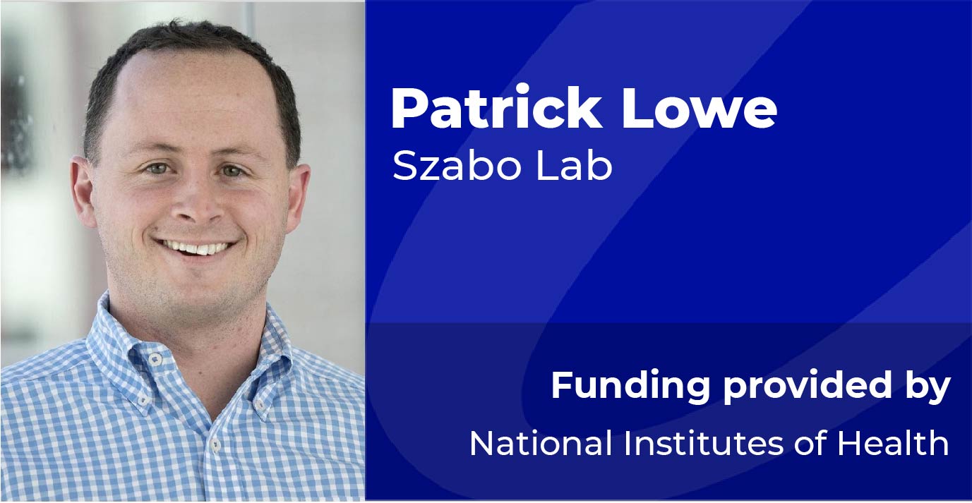
    The Role of Extracellular Vesicles in Alcohol-Induced Neuroinflammation

    The central nervous system is susceptible to many environmental insults and like many organs can be affected by alcohol. Alcohol impacts the brain in a variety of ways including short-term cognitive changes, development of dependence, memory deficits, neuronal loss and initiation of neuroinflammation. An emerging mechanism being studied in the field of central nervous system (CNS) inflammation, extracellular vesicle communication, has not yet been investigated in alcohol-related neuroinflammation and offers the potential for therapeutic intervention. Key components of alcohol-induced neuroinflammation, the cytokines IL-1β and HMGB1, are thought to be released from cells via extracellular vesicles. This study will explore the hypothesis that alcohol alters the release of extracellular vesicles within the CNS and that these vesicles contain content critical to the inflammatory process. Our Preliminary Data reveals that EVs are released by CNS cell types and can be taken up by unstimulated cells. First, we examined the effect of alcohol exposure on microglia and astrocytes in vitro and found that exosomes were stimulated for release at either 50 or 100mM alcohol. These findings were confirmed with western blot against exosome marker CD63 in the supernatant. Next, we used the membrane dye PKH26 to label membranes of microglia which were then stimulated to release EVs by alcohol. Those EVs were transferred to untreated/unlabeled cells and the dye was seen to incorporate in recipient cells, suggesting that those EVs were taken up by the untreated cells. Specific Aim 1 will investigate the effect of alcohol on extracellular vesicle release from primary mouse CNS cells (neurons, microglia or astrocytes) in single cell-type cultures in vitro. Nanoparticle tracking analysis will be used to measure released vesicles size, which will allow for quantification of the two types of released vesicles: exosomes (<150nm diameter) or microvesicles (150nm-1μm). Proinflammatory cytokines IL-1β and HMGB1 will then be measured in vesicles secreted from CNS cell types after alcohol exposure. These experiments will provide important knowledge regarding alcohol's impact on vesicle release as well as vesicle content. As extracellular vesicles are believed to transmit intercellular signals, Specific Aim 2 will explore the effect of transferring alcohol-induced vesicles onto naïve cells. First, extracellular vesicle uptake by primary CNS cell types will be measured. Next brain slices maintained in culture will be exposed to vesicles derived from alcohol-exposed cells and activation of inflammatory pathways will be examined. Finally, IL-1β or HMGB1 will be individually knocked down or overexpressed in CNS cell types and alcohol-induced vesicles will be transferred onto brain slices. These experiments will test the effect that alcohol-induced extracellular vesicles have on other cells, as well as the contribution made by cargo cytokines. Specific Aim 3 will elucidate the impact that alcohol-induced vesicles have on the brain in vivo. First, we will investigate the concentrations of EVs required for intracranial injection and uptake in the brain by using fluorescently-labeled vesicles. Next, vesicles will be stimulated in vitro from primary mouse CNS cells exposed to alcohol. After isolating those vesicles, they will be injected into the brains of naïve mice. Brain tissue will b examined for increases in immune cell activation and upregulation of inflammatory signals. This experiment will provide important information regarding the impact of extracellular vesicles on inflammation in vivo. The first year of this fellowship will be dedicated to quantifying and qualifying the vesicles released by CNS cells after alcohol exposure. Specific Aim 2 will be investigated in years two and three of the fellowship, while Specific Aim 3 will be completed in year three. The final two years of the fellowship will be dedicated to completing the clinical rotations for my MD training as well as any necessary follow up experiments needed for publishing this proposed work.

    Read more
  • Matthew.jpg

    Structure-based design of robust cross-genotypic NS3/4A protease inhibitors that avoid resistance

    Hepatitis C virus (HCV), a pathogen that infects over 150 million people worldwide, is the leading cause of chronic hepatitis, liver cirrhosis and hepatocellular carcinoma. HCV is a genetically diverse virus with 6 known genotypes with genotypes 1 and 3 being the most prevalent. This genetic diversity makes HCV infection difficult to treat. In the last few years, the advent of direct-acting antivirals (DAAs) has remarkably improved therapeutic options and treatment outcomes. However, despite highly potent inhibitors against multiple proteins, drug resistance is a major problem in all drug classes. Drug resistance is a loss of inhibitor potency while maintaining substrate processing. Though NS3/4A protease inhibitors are highly potent, they are not efficacious against all genotypes and are susceptible to drug resistance. Underlying differential inhibitor potency are the molecular mechanisms of drug resistance and genotypic differences. Elucidating these are key to developing protease inhibitors that avoid drug resistance and are effective against all HCV genotypes. Specifically most protease inhibitors in clinical development contain P2 moieties that contact unessential residues of the protease, which while increasing potency also increases their susceptibility to single site mutations that confer drug resistance. I hypothesize that protease inhibitors that avoid contact with these residues while leveraging contact with unexploited areas in the active site will result in inhibitors with enhanced potency and higher barriers to drug resistance. To investigate this hypothesis, using computational techniques, I will design a panel of novel protease inhibitors with extended P4 groups. I will then synthesize and enzymatically assay these protease inhibitors. Top leads will be co-crystalized with the protease and structurally analyzed to optimize the computational designs and initiate iterative rounds of inhibitor design. This project will provide molecular insights about the mechanisms of drug resistance as well as new strategies for the design of novel protease inhibitors for the effective treatment of HCV infection.

    Read more